Anatomical Evidence for Evolution
When one observes the anatomy of various animals, one finds an infinite number of similarities. reptiles, birds and amphibians.
• All mammals, reptiles, birds and amphibians – the tetrapods – have four limbs.
• All primates have five fingers in their extremities.
• All mammals share the same bones in their extremities.
• The beaks and wings of all birds are similar.
• The fins of all fish are similar.
• In general, species have organs in common.
The former suggests that present day species must share common ancestors from which they inherited those characteristics. Anatomical similarities between two species is evidence of their evolution from a common ancestor. Let us examine a few examples.
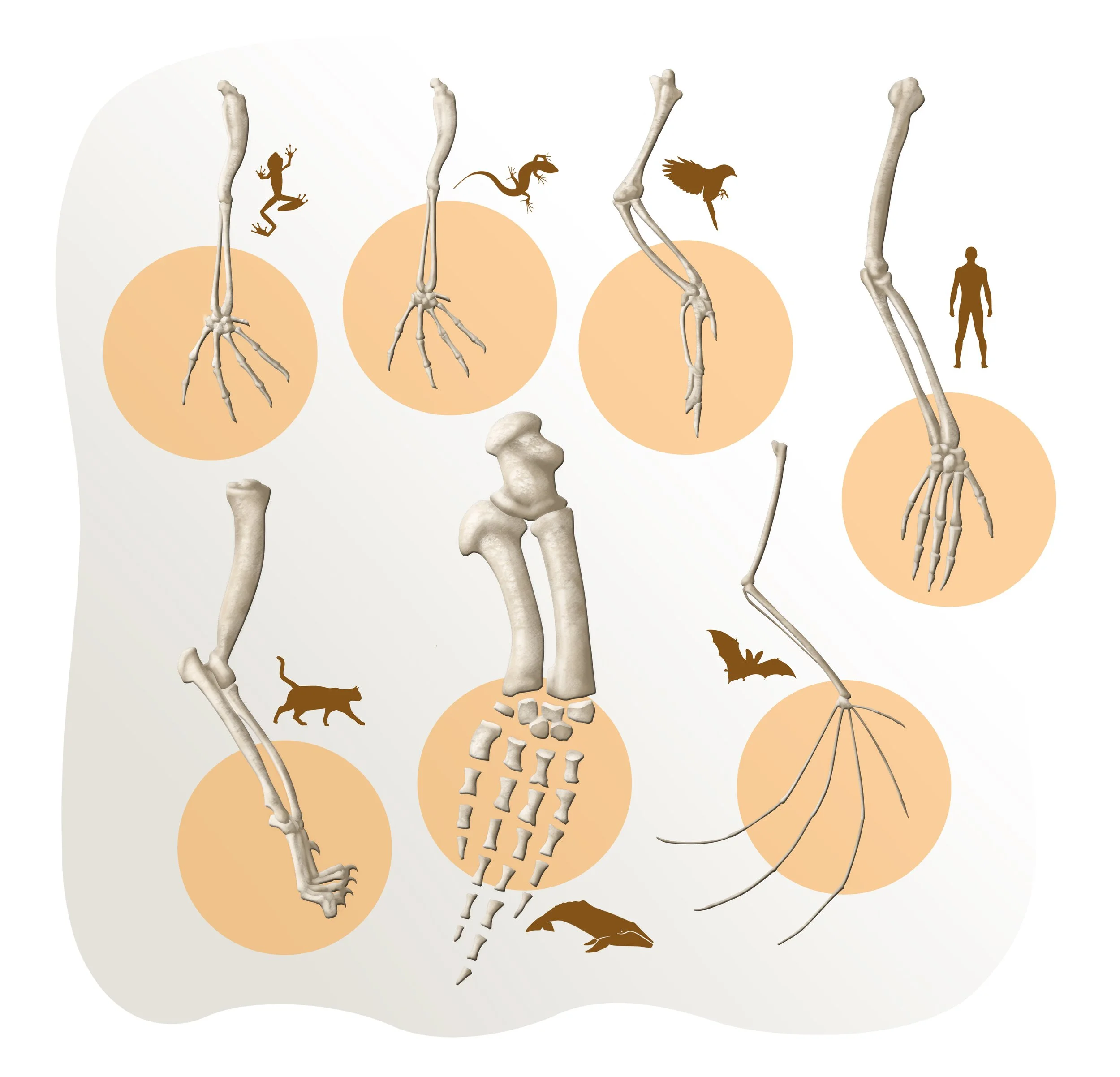
Upper limbs of (from top-left, clockwise) amphibian, reptile, bird, human, bat, cetacean and feline.
Embryonic development in vertebrates
All vertebrates begin their embryonic development in the same way, and in a similar way to the development of a fish. As they grow, different species begin to diverge. Some blood vessels, nerves or organs that appear at first, disappear suddenly, while others change their location or else transform themselves into something different. This puzzle only makes sense by thinking of evolution. The stages that follow one another during the embryonic development of a vertebrate, their sequence in fact, mirror the order in which their ancestors evolved.
For example, a lizard begins its development appearing like a fish embryo, it then resembles an amphibian embryo, to finally become a reptilian embryo. Mammals follow the same sequence, while ending in a mammalian embryonic stage. The explanation is that when a species evolves into another, its descendants inherit all the genes that form the ancestral structures. Later on, evolution takes advantage of the genetic material available and retools it, from which a new species can then appear.
A fish embryo
The Forelimb of Tetrapods
A tetrapod is an animal with a backbone that walks on two or four legs. The upper extremities of tetrapods (amphibians, mammals, birds, and reptiles) are made up of the same bones: the humerus, which articulates at the shoulder, the radius, and the ulna, which make up the lower arm, and many smaller bones in the wrist, hand, and fingers. The origin of the forelimb structure can be found in the fossil record.
The humerus, ulna, and radius are present in the fossil Eusthenopteron, a 380-million-year-old fish, slightly more ancient than tetrapods. Tiktaalik, a transition species between fish and tetrapods, possessed wrist, hand, and finger areas in its pectoral fins. The first fingers appeared in Acanthostega, the oldest tetrapod known to date, dating back 365 million years. The gradual change in this forelimb structure is evolutionary evidence that all tetrapods descended from a common ancestor.
Upper limbs of (from top-left, clockwise) amphibian, reptile, bird, human, bat, cetacean and feline.
The Vestigial Structures and Adaptations of Aquatic Mammals, the Cetaceans
Modern-day cetaceans (dolphins, whales, and porpoises) evolved from terrestrial mammals about 60 million years ago, when dinosaurs disappeared and left many empty ecological niches behind. Cetaceans carry their evolutionary history within their bodies; we can see the vestiges of their ancestors in their anatomy. When observing a whale skeleton in a natural history museum, one is struck by the presence of a few bones located at the midbody, separated from the spinal column. These vestigial structures are the remnants of two small hindlegs and a pelvis, all that is left of the lower extremities of its terrestrial ancestors. Vestigial structures are traits useful to the ancestor but no longer beneficial to the modern-day species. Since whales no longer have any use or function for these extremities, they have disappeared over generations. Snakes also have these vestigial structures. Known as pelvic spurs, they are made up of a tiny pelvis and two femurs, trace evidence of their tetrapod ancestors.
In addition, cetaceans evolved anatomical adaptations allowing them to see and hear underwater. Unlike other mammals, they can live in water and rise to the surface only to breathe. The cetaceans’ nasal cavity migrated to the upper part of the skull. In contrast, in terrestrial mammals, such as dogs, the nasal cavities are located at the end of their snouts, pointing forward and down. Dogs must lift their heads to breathe and avoid swallowing water when they swim. Dogs do not know what is happening underneath the water since their eyes and ears are above the surface. On the other hand, a dolphin can breathe calmly while maintaining their eyes open underwater, allowing them to react in case of danger.
Another feature pointing to the terrestrial origin of the whales appears during the development of the embryos of baleen whales, the toothless whales and finback whales. Instead of teeth, they have plates used to filter food from the water as it enters their mouths. It is surprising that mysticete embryos develop teeth that are reabsorbed and disappear by the end of gestation period.
Recreation of the skeleton of a Dorudon, an extinct genus of cetaceans,which still had small hind limbs.
A cetacean’s skull (left) has a nasal cavity (yellow) located above the eye socket (purple). In a dog’s skull, the nasal cavity is located at the end of the snout, below the eye sockets. (right).
The Vestiges of our Ancestors in the Human Body
The human body is full of the vestiges of its mammalian ancestors. These vestigial structures evolved in our ancestors because they were beneficial. The appendix, for example, is a small pouch located at the end of the small intestine. Its size varies in humans from 2 cm to 30 cm. Some individuals are born without one. Herbivorous animals, such as koalas, rabbits, and kangaroos, all have large appendices. The same is true for primates who eat leaves, such as lemurs, monkeys, and gorillas. The appendix allows them to ferment food by digesting the cellulose found in plants. Primates that feed on a few leaves, such as orangutans, have small appendices. We humans have a vestigial one, the remnants of an organ vital to our herbivorous ancestors.
Another example is the coccyx, fused vertebrae located at the base of the spinal column. The coccyx is a remnant of our ancestors’ tails, which disappeared around 20 million years ago. Interestingly, there have been cases of humans born with a small tail.
Goose bumps is another vestigial structure found in humans. When we get scared or cold, the small muscles at the base of our hair follicles contract. In humans, goose bumps serve no purpose, but in other mammals, they function as a thermal insulator. Goose bumps can also make an animal appear larger when threatened, like when a frightened cat’s fur stands on end or when a chimpanzee is about to fight.
Some human beings can move their ears. This action has no benefit since we depend more on sight than sound. Nevertheless, for a horse or a cat, it is a helpful way to localize the origin of a sound, an important strategy to avoid predators or locate one’s young. This movement surely benefitted our human ancestors, a fact reflected in members of our species today.
One last example is lanugo, the fine, soft hair covering a human fetus from around the sixth month of gestation to about one month before birth. Lanugo serves no purpose since the mother’s own body provides the fetus with the adequate temperature for its development. The only explanation for its existence is that the lanugo is a vestige of our primate past. Like goose bumps, it’s evidence that our apelike ancestors had fur. The fetuses of all monkeys and apes have lanugo, which develops into their fur. The fetuses of whales, like human fetuses, lose their lanugo before birth. It is a vestige of their hairy terrestrial ancestors.
We can conclude that the presence of vestigial structures is another line of evidence for evolution. We carry our evolutionary history, the traits of our ancient ancestors, within our own bodies.
Goosebumps
Darwin’s Comet Orchid
The comet, or star orchid, Angraecum sesquipedale, is a flower found on the island of Madagascar. Its most notable characteristic is the flower’s exceedingly long nectar spur, the long, green tubular extension that hangs off the flower and houses the nectar.
When Charles Darwin saw a specimen of this plant, he hypothesized that this flower was pollinated by a moth with an unusually long proboscis, one so long that it could reach into the depths of the nectar spur. A proboscis is a kind of trunk for suctioning out the nectar of a flower.
Nobody believed Darwin’s speculation at the time. It was not until 1903 that such an insect was found. The Madagascar moth, Xanthopan morgani praedicta, possesses a proboscis of just the right length. Scientists have video of the moth drinking nectar from Darwin’s comet orchid.
Darwin´s star orchid
The Wings of the Ostrich, the Penguin, and the Kiwi
Several species of birds have lost the ability to fly. These birds evolved in an environment where flight was not a necessary strategy for survival. The most common examples are ostriches, penguins, and kiwis.
Ostriches descended from flying birds. Evidence from both the fossil record and DNA confirms this hypothesis. Present-day ostriches evolved in an environment free of predators; they do not need to fly away to avoid being eaten. However, they still possess wings that now have several different functions. They help ostriches keep their balance, and the males use their wings to attract females. These birds can also use their wings to scare away enemies or protect their young.
Penguins are another interesting example of a flightless bird species. Their wings have become fins used to swim underwater at surprising speeds.
The most extreme case is that of the New Zealand kiwi. Kiwis have vestigial wings but don’t have any use for them. They are so tiny it’s hard to notice them beneath their plumage.
These flightless birds are located in different parts of the world yet possess very similar characteristics, providing us with evidence of common ancestry.
The wings of ostrich, penguin, and kiwi.
Three Types of Kidneys Develop in the Human Fetus
Human fetuses have three successive kidneys during gestation, with the first two degenerating entirely before birth. The first two kidneys resemble, in order, those of primitive jawless fish and amphibians.
The explanation is that human beings still possess the genes we inherited from our aquatic ancestors, and we go through the developmental stages that show organs resembling those of our ancestors.
The first type of kidney, the pronephric kidney, begins to form at three weeks of gestation. In lampreys and other primitive jawless fish, this organ filters wastes from the coelom (body cavity) and excretes them to the outside. The pronephric kidney does not function in humans and other mammalian embryos. It begins to disappear shortly after formation.
The second type of kidney is the mesonephric kidney. This kidney filters wastes from the blood and excretes them to the outside of the body via a pair of tubes called the mesonephric ducts, also known as Wolffian ducts. The mesonephric kidney develops into the adult kidney of fish and amphibians. This kidney functions for a few weeks in the human embryo but then disappears.
The third type of kidney is the metanephric kidney. It begins developing about five weeks into gestation and filters wastes from the blood. It excretes them to the outside through a pair of new tubes, the ureters. The metanephric kidney is the final adult kidney of reptiles, birds, and mammals.


